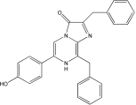
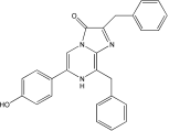
腔肠素h 天然腔肠素去羟基衍生物|Coelenterazine h
产品说明书
FAQ
COA
已发表文献
产品描述
腔肠素h(Coelenterazine h),天然腔肠素的去羟基衍生物,是海肾荧光素酶(Rluc)的作用底物,也是水母发光蛋白的辅因子,其发光强度比天然腔肠素高10倍以上,适用于报告基因分析。也是水母发光蛋白的辅助因子,可用于检测活细胞中钙离子浓度,基因报告分析,BRET(生物发光共振能量转移)研究,ELISA,HTS,以及组织或细胞中ROS水平的化学发光检测。
产品性质
|
英文别名(English synonym) |
2-Deoxycoelenterazine; CLZN-h; h-CTZ |
|
CAS号(CAS NO) |
50909-86-9 |
|
分子式(Formula) |
C26H21N3O2 |
|
分子量(Molecular weight) |
407.5 g/mol |
|
外观(Appearance) |
黄色固体 |
|
溶解性(Solubility) |
可溶于甲醇和乙醇,不可溶于DMSO |
|
纯度(Purity)(TLC) |
>99% |
|
结构(Structure) |
|
运输和保存方法
冰袋运输。粉末-20℃避光干燥保存,最好保存在惰性气体环境下,避免接触空气;长期保存于-70℃,有效期2年。
腔肠素h溶液的配制
对于所有进行荧光信号检测的试验建议溶液现用现配。建议可用乙醇或丙二醇配制成储存液(配制浓度可参考文献推荐),在使用时需将储存液用合适的缓冲液稀释成工作液。
注意事项
1)腔肠素h的干燥粉末在密封状态下较稳定,可避光保存于-20℃或更低温度。可通过在管内充入惰性气体(氮气或氩气)防止其氧化。
2)当使用多孔板进行荧光值的检测时,建议通过设置对照孔来消除由于腔肠素在工作液中不断被氧化所带来的误差。
3)该产品适用于体外生物发光检测;对于活体动物成像检测,建议使用 Ready To Use Coelenterazine h(见Yeasen 40907ES10)即用型腔肠素h。
4)不同种类的荧光素酶存在很大的区别。如腔肠素h和腔肠素F可作为海肾荧光素酶的底物,但对于Gaussia荧光素酶是无效的。
5)为了您的安全和健康,请穿实验服并戴一次性手套操作。
6)本产品仅作科研用途!
HB220826
Q: 腔肠素溶液如何配制?
A: (1)腔肠素溶液配制方式来源文献,仅供参考,具体工作浓度建议参考文献做梯度摸索最佳浓度。 母液:称取 1mg 腔肠素粉末直接溶解于 197µl 酸化的甲醇溶液( 100% 甲醇中含有 20µl/ml 3M 或者 6M HCl),配制成 12 mM (~5mg/ml,5×) 的腔肠素储存液。 工作液:如体外:10 的 5 次方的细胞数可用 10 μ M 。
Q:腔肠素的发光特性如何?
A: 体外:细胞孵育 10-15min 后,立即检测。 体内:可在注射后 10-15 min 内检测。
仅供参考,建议预实验建立荧光素酶动力学曲线,从而确定最高信号检测时间和信号平台期。
Q:是否能用于活体成像?
A: 一般用于体外,因为容易氧化,体内的话要注射和麻醉,时间久,体外检测快,不影响。体内的话推荐即用型。
Q:推荐仪器?多功能酶标仪能用吗?
A: 推荐仪器:具有生物化学发光检测模块。荧光素产生的光可以被光度计或闪烁计数器检测。常见的活体成像仪器:如 IVIS® Lumina 小动物活体成像系统,德国 Bruker 公司的 In-Vivo Xtreme 多模式小动物活体成像仪。多功能酶标仪:需要和仪器厂家确认是否具体生物化学发光检测模块。注:不能用荧光显微镜。
Q:说明书写到的配置储存液用的丙二醇是1,2-丙二醇吗?乙醇是无水乙醇吗?
A: 是的,丙二醇是1,2-丙二醇,乙醇是无水乙醇。
[1] Lin S, Han S, Cai X, et al. Structures of Gi-bound metabotropic glutamate receptors mGlu2 and mGlu4. Nature. 2021;594(7864):583-588. doi:10.1038/s41586-021-03495-2(IF:49.962)
[2] Ma S, Chen Y, Dai A, et al. Structural mechanism of calcium-mediated hormone recognition and Gβ interaction by the human melanocortin-1 receptor. Cell Res. 2021;31(10):1061-1071. doi:10.1038/s41422-021-00557-y(IF:25.617)
[3] Zhang H, Chen LN, Yang D, et al. Structural insights into ligand recognition and activation of the melanocortin-4 receptor. Cell Res. 2021;31(11):1163-1175. doi:10.1038/s41422-021-00552-3(IF:25.617)
[4] Shao Z, Shen Q, Yao B, et al. Identification and mechanism of G protein-biased ligands for chemokine receptor CCR1. Nat Chem Biol. 2022;18(3):264-271. doi:10.1038/s41589-021-00918-z(IF:15.040)
[5] Liu Q, Yang D, Zhuang Y, et al. Ligand recognition and G-protein coupling selectivity of cholecystokinin A receptor. Nat Chem Biol. 2021;17(12):1238-1244. doi:10.1038/s41589-021-00841-3(IF:15.040)
[6] Zhao F, Zhou Q, Cong Z, et al. Structural insights into multiplexed pharmacological actions of tirzepatide and peptide 20 at the GIP, GLP-1 or glucagon receptors. Nat Commun. 2022;13(1):1057. Published 2022 Feb 25. doi:10.1038/s41467-022-28683-0(IF:14.919)
[7] Zhou F, Zhang H, Cong Z, et al. Structural basis for activation of the growth hormone-releasing hormone receptor. Nat Commun. 2020;11(1):5205. Published 2020 Oct 15. doi:10.1038/s41467-020-18945-0(IF:12.121)
[8] Shao L, Chen Y, Zhang S, et al. Modulating effects of RAMPs on signaling profiles of the glucagon receptor family. Acta Pharm Sin B. 2022;12(2):637-650. doi:10.1016/j.apsb.2021.07.028(IF:11.614)
[9] Shao Z, Tan Y, Shen Q, et al. Molecular insights into ligand recognition and activation of chemokine receptors CCR2 and CCR3. Cell Discov. 2022;8(1):44. Published 2022 May 15. doi:10.1038/s41421-022-00403-4(IF:10.849)
[10] Zhao F, Zhang C, Zhou Q, et al. Structural insights into hormone recognition by the human glucose-dependent insulinotropic polypeptide receptor. Elife. 2021;10:e68719. Published 2021 Jul 13. doi:10.7554/eLife.68719(IF:8.146)
[11] Wang YZ, Yang DH, Wang MW. Signaling profiles in HEK 293T cells co-expressing GLP-1 and GIP receptors. Acta Pharmacol Sin. 2022;43(6):1453-1460. doi:10.1038/s41401-021-00758-6(IF:6.150)
[12] Wang J, Yang D, Cheng X, et al. Allosteric Modulators Enhancing GLP-1 Binding to GLP-1R via a Transmembrane Site. ACS Chem Biol. 2021;16(11):2444-2452. doi:10.1021/acschembio.1c00552(IF:5.100)
[13] Lin GY, Lin L, Cai XQ, et al. High-throughput screening campaign identifies a small molecule agonist of the relaxin family peptide receptor 4. Acta Pharmacol Sin. 2020;41(10):1328-1336. doi:10.1038/s41401-020-0390-x(IF:5.064)
[14] Darbalaei S, Yuliantie E, Dai A, et al. Evaluation of biased agonism mediated by dual agonists of the GLP-1 and glucagon receptors. Biochem Pharmacol. 2020;180:114150. doi:10.1016/j.bcp.2020.114150(IF:4.960)
[15] Yuliantie E, Darbalaei S, Dai A, et al. Pharmacological characterization of mono-, dual- and tri-peptidic agonists at GIP and GLP-1 receptors. Biochem Pharmacol. 2020;177:114001. doi:10.1016/j.bcp.2020.114001(IF:4.960)
[16] Sun L, Hao Y, Wang Z, Zeng Y. Constructing TC-1-GLUC-LMP2 Model Tumor Cells to Evaluate the Anti-Tumor Effects of LMP2-Related Vaccines. Viruses. 2018;10(4):145. Published 2018 Mar 23. doi:10.3390/v10040145(IF:3.761)


