细胞周期与细胞凋亡检测试剂盒说明书

细胞周期与细胞凋亡检测试剂盒说明书

产品详情

货号

规格

价格

78EA10010-100T

100T

询价

产品描述

细胞周期(cell cycle)是指连续分裂的细胞从一次分裂完成开始到下次分裂为止的全过程,分为间期和分裂期(M期),细胞间期又分为休眠期(G0期),DNA合成前期(G1期),DNA合成期(S期),DNA合成后期(G2期)整个周期可以表示为:G0-G1-S-G2-M

本公司生产的细胞周期检测是通过经典的碘化丙啶染色色 (Propidium iodide stainingPI staining)方法进行周期分析的检测试剂盒。PI是一种DNA的荧光染料,可以结合双链DNA产生荧光,其荧光强度与DNA的含量成正比。经碘化丙啶染色后假设G0/G1期细胞的荧光强度为1,那么含有双份基因组DNAG2/M期细胞的荧光强度的理论值为2,正在进行DNA复制的S期细胞的荧光强度为1-2之间。凋亡细胞由于细胞核发生浓缩以及发生DNA片段化导致部分基因组DNA片段在染色过程中丢失,因此凋亡细胞碘化丙啶染色后呈现明显的弱染,即荧光强度小于1,在流式检测的荧光图上出现所谓的sub-G1峰,即凋亡细胞峰。利用流式细胞仪进行荧光分析从而对DNA进行定量,实现细胞周期检测。

产品组成

本试剂盒常用于贴壁细胞或者悬浮细胞的培养,如果检测对象为组织样品,必须消化成单个细胞后在进行染色检测,规格为50T,每T可检测的样本的细胞数量在10-100万之间。

名称

规格

保存条件

染色缓冲液

25 mL

-20℃

碘化丙啶染色液(20X)

1.25 mL

-20℃

RNase A (50X)

0.5 mL

-20℃

运输与保存

冰袋运输,-20℃保存一年。

实验步骤

1. 细胞样品的准备:

a. 对于贴壁细胞:收集培养液上清,PBS润洗细胞一遍,胰酶消化细胞,加入前面收集的培养液终止消化,得到的细胞悬液1000g离心5min收集细胞沉淀备用。

b. 对于悬浮细胞:1000g左右离心3-5 min,沉淀细胞。

(如果细胞沉淀不充分,可以适当延长离心时间或稍稍加大离心力)

2.细胞固定:

1)向步骤1收集得到的沉淀中加入300 μL预冷的PBS,轻柔吹打混匀,轻柔涡旋的过程中滴加700 μL预冷的无水乙醇,最后于4℃固定30 min或更长时间。通常固定2 h或以上更能保证染色效果,固定12-24h可能效果更佳。

21000g左右离心5 min去除固定液,预冷PBS清洗一遍后再次离心,小心吸除上清,可以残留约50微升左右的PBS,以避免吸走细胞。轻轻弹击离心管底以适当分散细胞,避免细胞成团。

3. 细胞染色:

1)参考下表配制碘化丙啶染色液(配置好的碘化丙啶染色液4℃保存,宜当日内使用):

名称

1个样品

染色缓冲液

0.5 mL

碘化丙啶染色液(20X)

25 μL

RNase A (50X)

10 μL

总体积

0.535 mL

2)每个样品中加入0.5 mL的碘化丙啶染色液缓慢并充分重悬细胞沉淀, 37ºC避光孵育30min。随后可以4ºC或冰浴避光存放。染色完成后宜在24 h内完成流式检测。

4.流式检测分析:检测激发波长为488 nm处红色荧光,用流式细胞仪对DNA含量进行分析,确定细胞周期变化情况。

注意事项

(1)需自备PBS70%乙醇,整个过程避光操作。

2)为了您的安全和健康,请穿实验服并戴一次性手套。

3)本产品仅作科研用途!

细胞凋亡产品订购


细胞凋亡产品订购

简要描述:上海金畔生物科技有限公司细胞凋亡产品专业代理,具体产品信息欢迎电询:021-50837765

细胞凋亡产品订购

详细介绍

产品咨询

上海金畔生物科技有限公司细胞凋亡产品专业代理,具体产品信息欢迎电询:

细胞凋亡产品订购

细胞凋亡(apoptosis)一般是指机体细胞在发育过程中或在某些因素作用下,通过细胞内

基因及其产物的调控而发生的一种程序性细胞死亡(programmed cell death)。细胞凋亡对

胚胎发育及形态发生(morphogenesis)、组织内正常细胞群的稳定、机体的防御和免疫反

应、疾病或中毒时引起的细胞损伤、老化、肿瘤的发生进展起着重要作用,并具有潜在的治

疗意义。

细胞凋亡途径中各事件的发生是有时序性的,即各事件按先后顺序依次发生,zui终导致凋亡

小体的出现,细胞随着发生凋亡。典型特征为:细胞膜PS(磷脂酰丝氨酸)的外翻;线粒体

膜电位的丧失;细胞核凝缩和断裂。


一、PS(磷脂酰丝氨酸)在细胞外膜上的检测(Annexin V法):

        在正常的活细胞中,磷脂酰丝氨酸(phosphotidylserine,PS)位于细胞膜的内侧

,但在早期凋亡的细胞中,PS 从细胞膜的内侧翻转到细胞膜的表面,暴露在细胞外环境中

。Annexin-V(膜联蛋白-V)是一种分子量为35-36KD的Ca2+ 依赖性磷脂结合蛋白,能与PS

高亲和力结合。用荧光素(FITC、Alexa Fluor488等)标记的 Annexin-V 作为探针,利用

流式细胞仪或荧光显微镜可检测细胞凋亡的发生。另外,碘化丙啶(propidine iodide, PI)

是一种核酸染料,它不能透过完整的细胞膜,但在凋亡中晚期的细胞和死细胞,PI 能够透

过细胞膜而使细胞核红染。因此将 Annexin-V 与 PI 匹配使用,就可以将凋亡早晚期的细

胞以及死细胞区分开来。


产品编号 产品名称 规格
40302ES20 Annexin V-FITC/PI 细胞凋亡检测试剂盒 20T
40302ES50 Annexin V-FITC/PI 细胞凋亡检测试剂盒 50T
40302ES60 Annexin V-FITC/PI 细胞凋亡检测试剂盒 100T
40303ES20 Annexin V-EGFP/PI 细胞凋亡检测试剂盒 20T
40303ES50 Annexin V-EGFP/PI 细胞凋亡检测试剂盒 50T
40303ES60 Annexin V-EGFP/PI 细胞凋亡检测试剂盒 100T
40304ES20 Annexin V-Alexa Fluor647/PI 凋亡检测试剂盒 20T
40304ES50 Annexin V-Alexa Fluor647/PI 凋亡检测试剂盒 50T
40304ES60 Annexin V-Alexa Fluor647/PI 凋亡检测试剂盒 100T
40305ES20 Annexin V-Alexa Fluor488/PI 凋亡检测试剂盒 20T
40305ES50 Annexin V-Alexa Fluor488/PI 凋亡检测试剂盒 50T
40305ES60 Annexin V-Alexa Fluor488/PI 凋亡检测试剂盒 100T


上海金畔生物科技有限公司

ApexBio品牌代理

ApexBio

简要描述:

ApexBio Technology LLC 在生物研究肽和实验室试剂中。ApexBio在许多研究领域都具有广泛的产品线,例如:遗传学,细胞凋亡,肿瘤生物学等。ApexBio还提供各种定制服务,包括肽的合成,抗体的产生和发展分析。

ApexBio Technology LLC 在生物研究肽和实验室试剂中ApexBio在许多研究领域都具有广泛的产品线,例如:遗传学,细胞凋亡,肿瘤生物学等。ApexBio还提供各种定制服务,包括肽的合成,抗体的产生和发展分析。

ApexBio is a premier provider of peptides and lab reagents for biological research. We carry a broad product line in research areas such as Apoptosis, Epigenetics, Cancer Biology and so on. Apexbio also provides custom services including peptide synthesis, antibody production and assay development.

品牌

名称

1

A8882

5mg/10mg/25mg

ApexBio

Caspase-3/7 Inhibitor I

3

A1902

5mg/10mg/25mg

ApexBio

Bromodomain Inhibitor, (+)-JQ1

5

A2585

10mg/25mg/100mg

ApexBio

Q-VD-OPh hydrate

7

B1250

5mg/10mg/25mg

ApexBio

Atazanavir

9

A8206

5mg/50mg

ApexBio

UM 171

11

A3840

1mg/5mg/10mg

ApexBio

WEHI-539

13

A2606

1mg/5mg/20mg

ApexBio

Gap 26

15

A8802

5mg/50mg

ProSci品牌代理

ProSci

简要描述:

ProSci是世界的细胞凋亡抗体生产厂家和供应商,现有17000余种抗体,产品涵盖了细胞凋亡、信号转导、免疫、肿瘤、细胞生物、神经生物、传染病等领域。

ProSci是世界的细胞凋亡抗体生产厂家和供应商,现有17000余种抗体,产品涵盖了细胞凋亡、信号转导、免疫、肿瘤、细胞生物、神经生物、传染病等领域。


We are a leader among antibody development companies. Our research antibodies, custom antibody development services and leading-edge research programs help scientists, lab technicians, researchers and other technologists with reduced research costs, flexible & wide-ranging antibody services, online support and also experienced, first-hand knowledge of antibody uses as they relate to specific immunochemistry applications.


产品列表:


No.

品牌

货号

名称

规格

1

ProSci

28-711

1110033M05RIK Antibody

50 μg

2

ProSci

70-549

14-3-3 Antibody

0.05 mL

3

ProSci

79-205

4E BP1 (phospho Thr45) Antibody

0.1 mg

4

ProSci

XG-6146

Bovine Anti-Human IgG Antibody (H&L) Antibody

0.5 mg

5

ProSci

XR-9960

Goat Anti-Rabbit IgG (H&L) Antibody (FITC)

2 mg

6

ProSci

1325

Bladder Tumor Lysate

0.1 mg

7

ProSci

XBL-10333

Colon Diabetic Disease Lysate

0.1 mg

8

ProSci

92-393

Amiloride-binding protein 1 Recombinant Protein

0.05 mg

9

ProSci

PSI-1820

Alzheimers Disease gamma-Secretase Detection Set

1 Set

10

ProSci

PSI-1830

Matrillin Detection Set

1 Set

11

ProSci

1610

Mouse Anti-Chicken IgY Antibody [4A4A10] (biotin)

0.05 mg

12

ProSci

XR-9320

Rabbit Anti-Goat IgG (H&L) Antibody (HRP)

1 mg

13

ProSci

45-200

Activin receptor-like kinase 1 Antibody

0.1 mg

14

ProSci

91-991

B-Cell Linker Protein Recombinant Protein

0.05 mg

15

ProSci

70-504

AFP / Alpha-Fetoprotein Antibody

200 μl

Baicalin黄芩苷 细胞凋亡抑制剂/铁死亡抑制剂|CAS 21967-41-9

Baicalin黄芩苷 细胞凋亡抑制剂/铁死亡抑制剂|CAS 21967-41-9

产品说明书

FAQ

COA

已发表文献


Baicalin黄芩苷可显著减弱SH-SY5Y细胞中OGD/RO介导的细胞凋亡,可通过抑制细胞凋亡来减轻脑缺血再灌注损伤,还可以阻断脂氧合酶诱导的脂质过氧化,是一种铁死亡抑制剂(Ferroptosis抑制剂)。


产品信息

货号

54035ES25

规格

25 mg

价格

355


产品性质

英文别名(English Synonym)

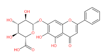
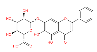
Baicalein 7-O-β-D-glucuronide; NSC-661431

中文名称 (Chinese Name)

黄芩苷

靶点(Target)

GABAA receptor; NF-κB

通路(Pathway)

NF-κB–NF-κB

CAS号(CAS NO.)

21967-41-9

分子式(Formula)

C21H18O11

分子量(Molecular Weight)

446.36

外观(Appearance)

粉末

纯度(Purity)

≥98%

溶解性(Solubility)

溶于DMSO

结构式(Structure)

Baicalin黄芩苷 细胞凋亡抑制剂/铁死亡抑制剂|CAS 21967-41-9


储存条件

-25~-15℃保存,有效期3年。避光干燥保存,避免反复冻融。


注意事项

1. 为了您的安全和健康,请穿实验服并戴一次性手套操作。

2. 粉末溶解前请先短暂离心,以保证产品全在管底。

3. 请勿吸入、吞咽或者直接接触皮肤和眼睛。

4. 本产品仅用于科研用途,禁止用于人身上。


使用浓度

【具体使用浓度请参考相关文献,并根据自身实验条件(如实验目的,细胞种类,培养特性等)进行摸索和优化。


使用方法(数据来自于公开发表的文献,仅供参考)

细胞实验(体外实验)

为了检测Baicalin黄芩苷对SH-SY5Y细胞缺氧和葡萄糖剥夺再灌注(OGD/RO)诱导损伤的神经保护作用,用黄芩苷 (1, 5, 25 μmol/L, 2 h)处理SH-SY5Y细胞OGD/RO组和对照组,MTT检测细胞活力,流式细胞术分析细胞凋亡,蛋白质印迹分析确定核因子(NF)-κB和N-甲基-天门冬氨酸受体-1 (NMDAR1)的表达。结果显示,黄芩苷可显著减弱SH-SY5Y细胞中OGD/RO介导的凋亡细胞死亡。低、中、高剂量组细胞凋亡率分别为12.1%、7.9%和5.4%。蛋白质印迹和实时荧光定量PCR分析显示,黄芩苷组半胱天冬酶-3表达与OGD/RO组相比显著降低(P<0.01)。此外,与OGD/RO组相比,黄芩苷组观察到NF-κB和NMDAR1的下调。与低剂量黄芩苷组相比,中、高剂量组显著下降。黄芩苷的预处理可通过抑制细胞凋亡来减轻脑缺血再灌注损伤。[1]


参考文献

[1] Zhou QB, et al. Pretreatment with baicalin attenuates hypoxia and glucose deprivation-induced injury in SH-SY5Y cells. Chin J Integr Med. 2016 Mar;22(3):201-6. doi: 10.1007/s11655-015-2326-8. Epub 2015 Dec 19. PMID: 26688183.

Ver.CN20230321

Baicalin黄芩苷 细胞凋亡抑制剂/铁死亡抑制剂|CAS 21967-41-9

暂无内容

Baicalin黄芩苷 细胞凋亡抑制剂/铁死亡抑制剂|CAS 21967-41-9

暂无内容

细胞周期检测试剂盒 细胞凋亡检测试剂盒|Cell Cycle and Apoptosis Analysis Kit

细胞周期检测试剂盒 细胞凋亡检测试剂盒|Cell Cycle and Apoptosis Analysis Kit

产品说明书

FAQ

COA

已发表文献

产品描述


细胞周期与细胞凋亡检测试剂盒(Cell Cycle and Apoptosis Analysis Kit)采用了经典的碘化丙啶染色(Propidium staining,即PI staining)方法对细胞周期与细胞凋亡进行分析。

碘化丙啶 (PropidiumPI) 是一种双链DNA荧光染料,其嵌入双链DNA后可以产生荧光,并且荧光强度和双链DNA的含量成正比。细胞内的DNAPI染色后,可以用流式细胞仪对细胞进行DNA含量测定,根据DNA含量的分布情况,进行细胞周期和细胞凋亡分析。

PI染色后,假设G0/G1期细胞的荧光强度为1,那么含有双份基因组DNAG2/M期细胞的荧光强度的理论值为2,正在进行DNA复制的S期细胞的荧光强度为1-2之间。凋亡细胞由于细胞核发生浓缩以及发生DNA片段化 (DNA fragmentation) 导致部分基因组DNA片断在染色过程中丢失,因此凋亡细胞PI染色后呈现明显的弱染,即荧光强度小于1,在流式检测的荧光图上出现所谓的sub-G1峰,即凋亡细胞峰。

细胞凋亡也可以用流式细胞仪观察细胞光散射的变化来检测。 细胞发生凋亡时,由于胞浆和染色质浓缩、核碎裂,产生凋亡小体,使细胞的光散射性质发生变化。凋亡前期,染色质皱缩,细胞密度增加,前向角光散射色显著降低。凋亡后期,细胞产生凋亡小体,前向角光散射和侧向角光散色都显著降低。

本试剂盒通常应用于贴壁或悬浮细胞的细胞周期与细胞凋亡检测。如果用于组织的细胞周期与细胞凋亡检测,则必须把组织消化成单细胞状态,才可以进行检测。


产品组分


编号

组分

产品编号/规格

40301ES5050 T

40301ES60(100 T)

40301-A

RNase A Solution

0.5 mL

1 mL

40301-B

PI Solution

0.5 mL

1 mL

40301-C

Staining Solution

25 mL

25 mL*2


运输与保存方法


运输:冰袋(wet ice)运输。

保存方法:-20℃避光保存,有效期为2年。

【注】如果需要在短时间内多次重复使用,可以于4℃避光保存2个月内有效。


注意事项


1)本试剂盒需要使用流式细胞仪进行检测。

2)细胞处理需轻柔,尽量避免人为的损伤细胞。

3)为防止不同批次细胞在实验时所处周期不同导致重复性差,可以在实验前进行细胞的同步化处理。实验细胞应处于对数

生长期,贴壁细胞一般在5080%汇合度时收集为宜。

4400目筛网过滤是用来将粘在一起的细胞团滤掉,留下单细胞,否则会出现人为的多倍体干扰。如果没有条件过滤,请在染色之前将细胞轻弹以分散,再进行染色。

5)荧光染料均存在淬灭问题,保存和使用过程中请尽量注意避光,以减缓荧光淬灭。

6)操作碘化丙啶时,应注意防护,保护眼睛、避免吸入。

7为了您的安全和健康,请穿实验服并戴一次性手套操作。

8) 本产品仅作科研用途!


使用方法


1)细胞样品制备:细胞数量控制在1×10⁵~1 × 10⁶个。

a)贴壁细胞:小心吸除细胞培养液,用胰酶消化细胞,制备成单细胞悬液。1000 g离心5 min沉淀细胞,弃上清,用1 mL预冷的PBS润洗细胞一次,离心收集细胞。

b)悬浮细胞:1000 g离心5 min,沉淀细胞,小心吸除上清。加入1 mL预冷的PBS,重悬细胞,再次离心收集细胞。

c)组织细胞:将组织块用剪刀剪成尽量小的小块后,用0.25%的胰酶消化0.5-1 h,经过200-400目筛网过滤得到单细胞悬液。1000 g离心5 min,沉淀细胞。加入约1 mL预冷的PBS,重悬细胞,再次离心沉淀细胞。如组织难以消化,可加入适量胶原酶。

2)细胞固定:细胞沉淀用1 mL预冷的70%乙醇轻轻混匀,4℃固定2 h以上或者过夜。接下来1000 g,离心5 min沉淀细

胞后,用1 mL预冷的PBS重悬。然后再次1000 g离心5 min沉淀细胞。

3)染色:

0.5 mL染色缓冲液(40301-C)中加入10 μL 碘化丙啶储液40301-B10 μLRNase A40301-A溶液,混匀待用。每个细胞样品加入0.5 mL配置好的碘化丙啶染色液,轻轻混匀重悬细胞。37℃避光孵育30 min就可以进行流式检测流式检测最好在5 h内完成。

【注】:配置好的PI染色液在短时间内可以4℃保存,宜当日使用。

4.流式检测和分析:细胞用400目筛网过滤,用流式细胞仪进行检测,在激发波长488 nm波长处检测,同时检测光散射情况。采用适当分析软件进行细胞DNA含量分析和光散射分析。

HB221122

Q:这款试剂盒是不是凋亡和周期均可以检测?

A:这款试剂盒是检测细胞周期的,细胞周期检测中细胞内的DNA被PI染色后,可以用流式细胞仪对细胞进行DNA含量测定,根据DNA含量的分布情况,进行细胞周期和细胞凋亡分析。


Q:Staining Solution能否使用PBS或DPBS代替?

A:可以


Q:流式周期结果如何分析?G1,S,G2-M数据分析。G2/G1的数据是有意义吗?

A:一般是G1期数据都基本没有变化,抑制细胞周期的药物处理就是G2/M期增加,S期减少。一般是看各个时期的比值


Q:流式周期固定的时候能否直接加70-75%的乙醇重悬细胞固定?

A:不可以,如果直接用70-75%的乙醇,会导致固定效果差或者出现细胞固定后无沉淀的现象。建议先用PBS重悬细胞后,再加入无水乙醇稀释到70-75%。


Q:做流式周期的时候,只出现单峰是什么原因?是不是你们的试剂有问题?

A:试剂是没有问题的,能出现峰说明试剂是可以和核酸结合的,只出现单峰或没有G2期,可能原因有以下几点:1.细胞状态,是不是细胞状态较差,建议调整细胞状态之后进行实验。2.细胞密度过密,如果细胞接种的时候过密会导致细胞接触抑制。3.原代细胞,有些原代细胞是没有增值能力的。4.圈门没圈好,建议正确圈门


Q:流式分析细胞周期,收集了10的6次方细胞,但细胞在乙醇固定之后,还可以看到细胞沉淀的,但PBS洗涤两次之后,就基本没什么细胞沉淀了,上机发现看不到细胞了。这是怎么回事啊?怎么解决这个问题啊?

A:1. 先用冷PBS悬浮细胞,充分悬浮,使细胞充分分散成单细胞。之后缓慢加入预冷的无水乙醇,终浓度为70-75%乙醇。不直接加75%乙醇的原因是:直接加入乙醇会导致细胞团聚的现象,很难重悬成单细胞。乙醇固定之后没有细胞沉淀。

2.很可能是洗细胞的过程中丢失了,解决办法有:采用尖底的离心管和水平离心机。离心后尽量用吸管吸取上清,不要倾倒;吸上清时残留1mm左右的水膜,不要吸完。离心的转速或时间可稍微增加一点儿。每次加溶液时,吸头最好不要接触液面;混匀时最好不要用吸头吹打,以免吸头挂壁带走部分细胞。


Q:试剂盒的 RNase A和 PI的浓度是多少,Staining Solution成分是什么?

A:RNase A和 PI的浓度都是1mg/ml,Staining Solution成分是一些盐溶液配方,配方不告知,用完了可以暂用PBS替代。


Q:周期的各个时期加起来不足100%是什么原因呢?

A:应该是没调好,因为流式是一个动态的过程,首位有点偏差是正常的,差的大可能是圈了一些黏连的细胞,需要去掉这些细胞,或者选择不同的拟合公式,可以调整圈门看看。

[1] Chen YY, Ge JY, Zhu SY, Shao ZM, Yu KD. Copy number amplification of ENSA promotes the progression of triple-negative breast cancer via cholesterol biosynthesis. Nat Commun. 2022;13(1):791. Published 2022 Feb 10. doi:10.1038/s41467-022-28452-z(IF:14.919)
[2] Zhao J, Lu P, Wan C, et al. Cell-fate transition and determination analysis of mouse male germ cells throughout development. Nat Commun. 2021;12(1):6839. Published 2021 Nov 25. doi:10.1038/s41467-021-27172-0(IF:14.919)
[3] Xia YK, Zeng YR, Zhang ML, et al. Tumor-derived neomorphic mutations in ASXL1 impairs the BAP1-ASXL1-FOXK1/K2 transcription network. Protein Cell. 2021;12(7):557-577. doi:10.1007/s13238-020-00754-2(IF:10.164)
[4] Luo Q, Lin L, Huang Q, et al. Dual stimuli-responsive dendronized prodrug derived from poly(oligo-(ethylene glycol) methacrylate)-based copolymers for enhanced anti-cancer therapeutic effect. Acta Biomater. 2022;143:320-332. doi:10.1016/j.actbio.2022.02.033(IF:8.947)
[5] Zhou Z, Fan T, Yan Y, et al. One stone with two birds: Phytic acid-capped platinum nanoparticles for targeted combination therapy of bone tumors. Biomaterials. 2019;194:130-138. doi:10.1016/j.biomaterials.2018.12.024(IF:8.806)
[6] Du P, Wang T, Wang H, Yang M, Yin H. Mucin-fused myeloid-derived growth factor (MYDGF164) exhibits a prolonged serum half-life and alleviates fibrosis in chronic kidney disease. Br J Pharmacol. 2022;179(16):4136-4156. doi:10.1111/bph.15851(IF:8.740)
[7] Wang J, Du X, Wang X, et al. Tumor-derived miR-378a-3p-containing extracellular vesicles promote osteolysis by activating the Dyrk1a/Nfatc1/Angptl2 axis for bone metastasis. Cancer Lett. 2022;526:76-90. doi:10.1016/j.canlet.2021.11.017(IF:8.679)
[8] Chen L, Su Y, Yin B, et al. LARP6 Regulates Keloid Fibroblast Proliferation, Invasion, and Ability to Synthesize Collagen [published online ahead of print, 2022 Feb 15]. J Invest Dermatol. 2022;S0022-202X(22)00116-6. doi:10.1016/j.jid.2022.01.028(IF:8.551)
[9] Chen W, Song J, Liu S, et al. USP9X promotes apoptosis in cholangiocarcinoma by modulation expression of KIF1Bβ via deubiquitinating EGLN3. J Biomed Sci. 2021;28(1):44. Published 2021 Jun 10. doi:10.1186/s12929-021-00738-2(IF:8.410)
[10] Duan Z , Luo Q , Gu L , et al. A co-delivery nanoplatform for a lignan-derived compound and perfluorocarbon tuning IL-25 secretion and the oxygen level in tumor microenvironments for meliorative tumor radiotherapy. Nanoscale. 2021;13(32):13681-13692. doi:10.1039/d1nr03738b(IF:7.790)
[11] Lu T, Lu H, Duan Z, et al. Discovery of High-Affinity Inhibitors of the BPTF Bromodomain. J Med Chem. 2021;64(16):12075-12088. doi:10.1021/acs.jmedchem.1c00721(IF:7.446)
[12] Zhang L, Zhao J, Dong J, Liu Y, Xuan K, Liu W. GSK3β rephosphorylation rescues ALPL deficiency-induced impairment of odontoblastic differentiation of DPSCs. Stem Cell Res Ther. 2021;12(1):225. Published 2021 Apr 6. doi:10.1186/s13287-021-02235-7(IF:6.832)
[13] Yu H, Yang X, Xiao X, et al. Human Adipose Mesenchymal Stem Cell-derived Exosomes Protect Mice from DSS-Induced Inflammatory Bowel Disease by Promoting Intestinal-stem-cell and Epithelial Regeneration. Aging Dis. 2021;12(6):1423-1437. Published 2021 Sep 1. doi:10.14336/AD.2021.0601(IF:6.745)
[14] Wang Y, Zhao M, Li W, et al. BMSC-Derived Small Extracellular Vesicles Induce Cartilage Reconstruction of Temporomandibular Joint Osteoarthritis via Autotaxin-YAP Signaling Axis. Front Cell Dev Biol. 2021;9:656153. Published 2021 Apr 1. doi:10.3389/fcell.2021.656153(IF:6.684)
[15] Zhang Y, Chen G, Zhuang X, Guo M. Inhibition of Growth of Colon Tumors and Proliferation of HT-29 Cells by Warburgia ugandensis Extract through Mediating G0/G1 Cell Cycle Arrest, Cell Apoptosis, and Intracellular ROS Generation. Oxid Med Cell Longev. 2021;2021:8807676. Published 2021 Dec 29. doi:10.1155/2021/8807676(IF:6.543)
[16] Pan X, Liu N, Liu Y, et al. Design, synthesis, and biological evaluation of trizole-based heteroaromatic derivatives as Bcr-Abl kinase inhibitors. Eur J Med Chem. 2022;238:114425. doi:10.1016/j.ejmech.2022.114425(IF:6.514)
[17] Wang CJ, Guo X, Zhai RQ, et al. Discovery of penipanoid C-inspired 2-(3,4,5-trimethoxybenzoyl)quinazolin-4(3H)-one derivatives as potential anticancer agents by inhibiting cell proliferation and inducing apoptosis in hepatocellular carcinoma cells. Eur J Med Chem. 2021;224:113671. doi:10.1016/j.ejmech.2021.113671(IF:6.514)
[18] Qin X, Dang W, Yang X, Wang K, Kebreab E, Lyu L. Neddylation inactivation affects cell cycle and apoptosis in sheep follicular granulosa cells [published online ahead of print, 2022 May 16]. J Cell Physiol. 2022;10.1002/jcp.30777. doi:10.1002/jcp.30777(IF:6.384)
[19] Zhang YL, Chen GL, Liu Y, Zhuang XC, Guo MQ. Stimulation of ROS Generation by Extract of Warburgia ugandensis Leading to G0/G1 Cell Cycle Arrest and Antiproliferation in A549 Cells. Antioxidants (Basel). 2021;10(10):1559. Published 2021 Sep 30. doi:10.3390/antiox10101559(IF:6.313)
[20] Xue J, Li S, Shi P, et al. The ETS Inhibitor YK-4-279 Suppresses Thyroid Cancer Progression Independent of TERT Promoter Mutations. Front Oncol. 2021;11:649323. Published 2021 Jun 16. doi:10.3389/fonc.2021.649323(IF:6.244)
[21] Han L, Wu Y, Liu F, Zhang H. eIF4A1 Inhibitor Suppresses Hyperactive mTOR-Associated Tumors by Inducing Necroptosis and G2/M Arrest. Int J Mol Sci. 2022;23(13):6932. Published 2022 Jun 22. doi:10.3390/ijms23136932(IF:5.924)
[22] Xu X, Yuan X, Ni J, et al. MAGI2-AS3 inhibits breast cancer by downregulating DNA methylation of MAGI2. J Cell Physiol. 2021;236(2):1116-1130. doi:10.1002/jcp.29922(IF:5.546)
[23] Chen X, Lin S, Lin Y, et al. BRAF-activated WT1 contributes to cancer growth and regulates autophagy and apoptosis in papillary thyroid carcinoma. J Transl Med. 2022;20(1):79. Published 2022 Feb 5. doi:10.1186/s12967-022-03260-7(IF:5.531)
[24] Sun W, Sun F, Meng J, et al. Design, semi-synthesis and bioactivity evaluation of novel podophyllotoxin derivatives as potent anti-tumor agents. Bioorg Chem. 2022;126:105906. doi:10.1016/j.bioorg.2022.105906(IF:5.275)
[25] Ma Y, Yang X, Han H, et al. Design, synthesis and biological evaluation of anilide (dicarboxylic acid) shikonin esters as antitumor agents through targeting PI3K/Akt/mTOR signaling pathway. Bioorg Chem. 2021;111:104872. doi:10.1016/j.bioorg.2021.104872(IF:5.275)
[26] Luo Z , Xue K , Zhang X , et al. Thermogelling chitosan-based polymers for the treatment of oral mucosa ulcers. Biomater Sci. 2020;8(5):1364-1379. doi:10.1039/c9bm01754b(IF:5.251)
[27] Sun C, Wei J, Long Z, et al. Spindle pole body component 24 homolog potentiates tumor progression via regulation of SRY-box transcription factor 2 in clear cell renal cell carcinoma. FASEB J. 2022;36(2):e22086. doi:10.1096/fj.202101310R(IF:5.192)
[28] Quan JH, Gao FF, Ismail HAHA, et al. Silver Nanoparticle-Induced Apoptosis in ARPE-19 Cells Is Inhibited by Toxoplasma gondii Pre-Infection Through Suppression of NOX4-Dependent ROS Generation. Int J Nanomedicine. 2020;15:3695-3716. Published 2020 May 26. doi:10.2147/IJN.S244785(IF:5.115)
[29] Bian L, Meng Y, Zhang M, et al. ATM Expression Is Elevated in Established Radiation-Resistant Breast Cancer Cells and Improves DNA Repair Efficiency. Int J Biol Sci. 2020;16(7):1096-1106. Published 2020 Feb 4. doi:10.7150/ijbs.41246(IF:4.858)
[30] Liu J, Tan F, Liu X, Yi R, Zhao X. Exploring the Antioxidant Effects and Periodic Regulation of Cancer Cells by Polyphenols Produced by the Fermentation of Grape Skin by Lactobacillus plantarum KFY02. Biomolecules. 2019;9(10):575. Published 2019 Oct 6. doi:10.3390/biom9100575(IF:4.694)
[31] Chen X, Tang Y, Yan J, Li L, Jiang L, Chen Y. Circ_0062270 upregulates EPHA2 to facilitate melanoma progression via sponging miR-331-3p. J Dermatol Sci. 2021;103(3):176-182. doi:10.1016/j.jdermsci.2021.08.005(IF:4.563)
[32] Xu A, Wang Q, Lin T. Low-Frequency Magnetic Fields (LF-MFs) Inhibit Proliferation by Triggering Apoptosis and Altering Cell Cycle Distribution in Breast Cancer Cells. Int J Mol Sci. 2020;21(8):2952. Published 2020 Apr 22. doi:10.3390/ijms21082952(IF:4.556)
[33] Wang J, Teng F, Chai H, Zhang C, Liang X, Yang Y. GNA14 stimulation of KLF7 promotes malignant growth of endometrial cancer through upregulation of HAS2. BMC Cancer. 2021;21(1):456. Published 2021 Apr 23. doi:10.1186/s12885-021-08202-y(IF:4.430)
[34] Sang L, Wu X, Yan T, et al. The m6A RNA methyltransferase METTL3/METTL14 promotes leukemogenesis through the mdm2/p53 pathway in acute myeloid leukemia. J Cancer. 2022;13(3):1019-1030. Published 2022 Jan 4. doi:10.7150/jca.60381(IF:4.207)
[35] Cao Y, Xie X, Li M, Gao Y. CircHIPK2 Contributes to DDP Resistance and Malignant Behaviors of DDP-Resistant Ovarian Cancer Cells Both in vitro and in vivo Through circHIPK2/miR-338-3p/CHTOP ceRNA Pathway. Onco Targets Ther. 2021;14:3151-3165. Published 2021 May 13. doi:10.2147/OTT.S291823(IF:4.147)
[36] Liu Z, Li Y, Li X, et al. Overexpression of YBX1 Promotes Pancreatic Ductal Adenocarcinoma Growth via the GSK3B/Cyclin D1/Cyclin E1 Pathway. Mol Ther Oncolytics. 2020;17:21-30. Published 2020 Mar 29. doi:10.1016/j.omto.2020.03.006(IF:4.115)
[37] Jiang L, Wang Y, Liu G, et al. C-Phycocyanin exerts anti-cancer effects via the MAPK signaling pathway in MDA-MB-231 cells. Cancer Cell Int. 2018;18:12. Published 2018 Jan 25. doi:10.1186/s12935-018-0511-5(IF:3.960)
[38] Wang RQ, He FZ, Meng Q, et al. Tribbles pseudokinase 3 (TRIB3) contributes to the progression of hepatocellular carcinoma by activating the mitogen-activated protein kinase pathway. Ann Transl Med. 2021;9(15):1253. doi:10.21037/atm-21-2820(IF:3.932)
[39] Li MT, Pi XX, Cai XL, et al. Ferredoxin reductase regulates proliferation, differentiation, cell cycle and lipogenesis but not apoptosis in SZ95 sebocytes. Exp Cell Res. 2021;405(2):112680. doi:10.1016/j.yexcr.2021.112680(IF:3.905)
[40] Wu H, Chen L, Zhu F, Han X, Sun L, Chen K. The Cytotoxicity Effect of Resveratrol: Cell Cycle Arrest and Induced Apoptosis of Breast Cancer 4T1 Cells. Toxins (Basel). 2019;11(12):731. Published 2019 Dec 13. doi:10.3390/toxins11120731(IF:3.895)
[41] Wang X, Zhang R, Wu T, et al. Successive treatment with naltrexone induces epithelial-mesenchymal transition and facilitates the malignant biological behaviors of bladder cancer cells. Acta Biochim Biophys Sin (Shanghai). 2021;53(2):238-248. doi:10.1093/abbs/gmaa169(IF:3.848)
[42] Ye F, Zhang W, Ye X, Jin J, Lv Z, Luo C. Identification of Selective, Cell Active Inhibitors of Protein Arginine Methyltransferase 5 through Structure-Based Virtual Screening and Biological Assays. J Chem Inf Model. 2018;58(5):1066-1073. doi:10.1021/acs.jcim.8b00050(IF:3.804)
[43] Shi G, Wang TT, Quan JH, et al. Sox9 facilitates proliferation, differentiation and lipogenesis in primary cultured human sebocytes. J Dermatol Sci. 2017;85(1):44-50. doi:10.1016/j.jdermsci.2016.10.005(IF:3.739)
[44] Liu J, Tan F, Liu X, Yi R, Zhao X. Grape skin fermentation by Lactobacillus fermentum CQPC04 has anti-oxidative effects on human embryonic kidney cells and apoptosis-promoting effects on human hepatoma cells. RSC Adv. 2020;10(8):4607-4620. Published 2020 Jan 29. doi:10.1039/c9ra09863a(IF:3.119)
[45] Liu J, Jiang C, Ma X, Feng L, Wang J. Notoginsenoside Fc Accelerates Reendothelialization following Vascular Injury in Diabetic Rats by Promoting Endothelial Cell Autophagy. J Diabetes Res. 2019;2019:9696521. Published 2019 Sep 3. doi:10.1155/2019/9696521(IF:3.040)
[46] Feng Q, Wang D, Guo P, Zhang Z, Feng J. Long non-coding RNA HOTAIR promotes the progression of synovial sarcoma through microRNA-126/stromal cell-derived factor-1 regulation. Oncol Lett. 2021;21(6):444. doi:10.3892/ol.2021.12705(IF:2.967)
[47] Chai B, Guo Y, Zhu N, et al. Pleckstrin 2 is a potential drug target for colorectal carcinoma with activation of APC/β‑catenin. Mol Med Rep. 2021;24(6):862. doi:10.3892/mmr.2021.12502(IF:2.952)
[48] An J, Wang H, Ma X, et al. Musk ketone induces apoptosis of gastric cancer cells via downregulation of sorbin and SH3 domain containing 2. Mol Med Rep. 2021;23(6):450. doi:10.3892/mmr.2021.12089(IF:2.952)
[49] Hassan RN, Luo H, Jiang W. Effects of Nicotinamide on Cervical Cancer-Derived Fibroblasts: Evidence for Therapeutic Potential. Cancer Manag Res. 2020;12:1089-1100. Published 2020 Feb 12. doi:10.2147/CMAR.S229395(IF:2.886)
[50] Li J, Jiang S, Chen Y, et al. Benzene metabolite hydroquinone induces apoptosis of bone marrow mononuclear cells through inhibition of β-catenin signaling. Toxicol In Vitro. 2018;46:361-369. doi:10.1016/j.tiv.2017.08.018(IF:2.866)
[51] Fang G, Wu Y, Zhang X. CircASXL1 knockdown represses the progression of colorectal cancer by downregulating GRIK3 expression by sponging miR-1205. World J Surg Oncol. 2021;19(1):176. Published 2021 Jun 14. doi:10.1186/s12957-021-02275-6(IF:2.754)
[52] Zhang Y, Sun C, Xiao G, Gu Y. Host defense peptide Hymenochirin-1B induces lung cancer cell apoptosis and cell cycle arrest through the mitochondrial pathway. Biochem Biophys Res Commun. 2019;512(2):269-275. doi:10.1016/j.bbrc.2019.03.029(IF:2.705)
[53] Liu XH, Zou J, Li YJ, et al. Isosteroidal alkaloids from Fritillaria hupehensis Hsiao et K.C.Hsia: Synthesis and biological evaluation of alkaloid derivatives as potential cytotoxic agents. Steroids. 2021;176:108929. doi:10.1016/j.steroids.2021.108929(IF:2.668)
[54] Shi G, Liao PY, Cai XL, et al. FoxO1 enhances differentiation and apoptosis in human primary keratinocytes. Exp Dermatol. 2018;27(11):1254-1260. doi:10.1111/exd.13775(IF:2.608)
[55] Zhang MF, Cai XL, Jing KP, et al. Differentiation Model Establishment and Differentiation-Related Protein Screening in Primary Cultured Human Sebocytes. Biomed Res Int. 2018;2018:7174561. Published 2018 Apr 5. doi:10.1155/2018/7174561(IF:2.583)
[56] Cheng YY, Yang X, Gao X, Song SX, Yang MF, Xie FM. LGR6 promotes glioblastoma malignancy and chemoresistance by activating the Akt signaling pathway. Exp Ther Med. 2021;22(6):1364. doi:10.3892/etm.2021.10798(IF:2.447)
[57] Chai M, Jiang M, Gu C, et al. Osteogenically differentiated mesenchymal stem cells promote the apoptosis of human umbilical vein endothelial cells in vitro [published online ahead of print, 2021 Oct 25]. Biotechnol Appl Biochem. 2021;10.1002/bab.2274. doi:10.1002/bab.2274(IF:2.431)
[58] Cheng Y, Yin Z, Jiang F, Xu J, Chen H, Gu Q. Two new lignans from the aerial parts of Saururus chinensis with cytotoxicity toward nasopharyngeal carcinoma. Fitoterapia. 2020;141:104344. doi:10.1016/j.fitote.2019.104344(IF:2.431)
[59] Lin C, Sun L, Huang S, Weng X, Wu Z. STC2 Is a Potential Prognostic Biomarker for Pancreatic Cancer and Promotes Migration and Invasion by Inducing Epithelial-Mesenchymal Transition. Biomed Res Int. 2019;2019:8042489. Published 2019 Jul 15. doi:10.1155/2019/8042489(IF:2.197)
[60] Wang B, Zhang XL, Li CX, Liu NN, Hu M, Gong ZC. ANLN promotes carcinogenesis in oral cancer by regulating the PI3K/mTOR signaling pathway. Head Face Med. 2021;17(1):18. Published 2021 Jun 3. doi:10.1186/s13005-021-00269-z(IF:2.151)
[61] Chen X, Xing M. Effects of 5-Aza-2'-deoxycytidine on hormone secretion and epigenetic regulation in sika deer ovarian granulosa cells. Reprod Domest Anim. 2021;56(2):360-369. doi:10.1111/rda.13873(IF:1.641)
[62] Wu S, Yang S, Qu H. circ_CHFR regulates ox-LDL-mediated cell proliferation, apoptosis, and EndoMT by miR-15a-5p/EGFR axis in human brain microvessel endothelial cells. Open Life Sci. 2021;16(1):1053-1063. Published 2021 Sep 29. doi:10.1515/biol-2021-0082(IF:0.938)

依托泊甙(Etoposide 100 mM) 抗肿瘤试剂 凋亡诱导剂 拓扑异构酶II抑制剂

依托泊甙(Etoposide 100 mM) 抗肿瘤试剂 凋亡诱导剂 拓扑异构酶II抑制剂

产品说明书

FAQ

COA

已发表文献

 产品描述

依托泊甙,是一种抗肿瘤试剂,作为一种凋亡诱导剂,依托泊甙是一种拓扑异构酶II抑制剂(IC50=59.2 μM),常用工作浓度5-50 µM


产品性质

英文别名(English Synonym

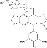
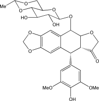
4'-Demethylepipodophyllotoxin; EPEG1; VP-16; VP-16213; NK 171; NSC-141540; 9-(4,6-O-Ethylidene-b-D-Glucopyranoside)

CAS号(CAS NO.

33419-42-0

分子式(Formula

C29H32O13

分子量(Molecular Weight

588.56

纯度(Purity

≥97%

溶解性(Solubility

溶于DMSO

结构式(Structure

依托泊甙(Etoposide 100 mM) 抗肿瘤试剂 凋亡诱导剂 拓扑异构酶II抑制剂 


运输和保存方法

冰袋运输。-20℃保存,冻干粉形式的Etoposide可以稳定保存24个月;液体形式的Etoposide建议在6个月内使用完,分装避免反复冻融。 


注意事项

1为了您的安全和健康,请穿实验服并戴一次性手套操作。

2本产品仅作科研用途!

HB221011

Q:可以用来哪些肿瘤细胞凋亡呢?

A:小细胞肺癌,恶性淋巴瘤,恶性生殖细胞瘤,白血病,对神经母细胞瘤,横纹肌肉瘤,卵巢癌,非小细胞肺癌,胃癌和食管癌等

[1] Li Y, Sun H, Zhang C, et al. Identification of translationally controlled tumor protein in promotion of DNA homologous recombination repair in cancer cells by affinity proteomics. Oncogene. 2017;36(50):6839-6849. doi:10.1038/onc.2017.289(IF:7.519)

BioVision品牌代理

BioVision 品牌介绍

简要描述:

BioVision. Inc. 位于美国加州的Palo Alto,是世界上专著于凋亡和细胞信号研究的公司。其产品质优、价廉、包装使用方便。BioVision致力于为研究者们提供好、研究工具而不断扩大产品及服务质量。该公司提供用于检测早、中、晚期凋亡的试剂,可以检测凋亡发生的不同区域,包括:线粒体、胞浆、胞膜及胞核。

BioVision. Inc. 位于美国加州的Palo Alto,是世界上专著于凋亡和细胞信号研究的公司。其产品质优、价廉、包装使用方便。BioVision致力于为研究者们提供好、研究工具而不断扩大产品及服务质量。该公司提供用于检测早、中、晚期凋亡的试剂,可以检测凋亡发生的不同区域,包括:线粒体、胞浆、胞膜及胞核。


Biovision was founded in 1998 by Swiss World Food Prize recipient Dr. Hans Rudolf Herren, with the aim of sustainably improving life for people in Africa while conserving the environment as the basis for all life.


In the 1980s, the world renowned entomologist Hans Rudolf Herren saved millions of people in Africa from starving to death by devising organic control methods for a devastating cassava pest. He was awarded the World Food Prize in 1995 for his work – he is the first, and so far only, Swiss person to have received it.


Biovision Foundation combats hunger and poverty, and is committed to the dissemination and application of ecological methods that sustainably improve living conditions in Africa whilst also conserving the environment. Biovision renders ‚help for self-help‘ and promotes ecological thought and action in both North and South.


In 2012, Biovision became the first Swiss Foundation to be granted General Consultative Status at ECOSOC of the United Nations. This allows Biovision to take part in UN Conferences, participate in official discussions and organize side events in order to argue its case.


In 2013, Biovision and its founder Hans Rudolf Herren won the Right Livelihood Award, also known as the Alternative Nobel Prize.


Biovision Foundation is a charitable organization in Switzerland (ZEWO certified) and has a legal opinion for 501(c)(3) public charity equivalency in the United States.

产品列表:

BioVision

K101-100

Annexin V-FITC Apoptosis Kit

100 assays

BioVision

K101-25

Annexin V-FITC Apoptosis Kit

25 assays

BioVision

K101-400

Annexin V-FITC Apoptosis Kit

400 assays

BioVision

K102-100

Annexin V-Cy3 Apoptosis Kit

100 assays

BioVision

K102-25

Annexin V-Cy3 Apoptosis Kit

25 assays

BioVision

K102-400

Annexin V-Cy3 Apoptosis Kit

400 assays

BioVision

K103-100

Annexin V-Cy5 Apoptosis Kit

100 assays

BioVision

K103-25

Annexin V-Cy5 Apoptosis Kit

25 assays

BioVision

K103-400

Annexin V-Cy5 Apoptosis Kit

400 assays

BioVision

K104-100

Annexin V-EGFP Apoptosis Kit

100 assays

BioVision

K104-25

Annexin V-EGFP Apoptosis Kit

25 assays

BioVision

K104-400

Annexin V-EGFP Apoptosis Kit

400 assays

BioVision

K105-100

Caspase-3 Fluorometric Assay Kit

100 assays

BioVision

K105-200

Caspase-3 Fluorometric Assay Kit

200 assays

BioVision

K105-25

Caspase-3 Fluorometric Assay Kit

25 assays

BioVision

K105-400

Caspase-3 Fluorometric Assay Kit

400 assays

BioVision

K106-100

Caspase-3 Colorimetric Assay Kit

100 assays

BioVision

K106-200

Caspase-3 Colorimetric Assay Kit

200 assays

BioVision

K106-25

Caspase-3 Colorimetric Assay Kit

25 assays

BioVision

K106-400

Caspase-3 Colorimetric Assay Kit

400 assays

BioVision

K109-100

Annexin V-Biotin Apoptosis Kit

100 assays

BioVision

K109-25

Annexin V-Biotin Apoptosis Kit

25 assays

BioVision

K109-400

Annexin V-Biotin Apoptosis Kit

400 assays

BioVision

K110-100

Caspase-1 Fluorometric Assay Kit

100 assays

BioVision

K110-200

Caspase-1 Fluorometric Assay Kit

200 assays

BioVision

K110-25

Caspase-1 Fluorometric Assay Kit

25 assays

BioVision

K110-400

Caspase-1 Fluorometric Assay Kit

400 assays

BioVision

K111-100

Caspase-1 Colorimetric Assay Kit

100 assays

BioVision

K111-200

Caspase-1 Colorimetric Assay Kit

200 assays

BioVision

K111-25

Caspase-1 Colorimetric Assay Kit

25 assays

BioVision

K111-400

Caspase-1 Colorimetric Assay Kit

400 assays

BioVision

K112-100

Caspase-8 Fluorometric Assay Kit

100 assays

Smac凋亡蛋白抑制剂|AT-406(SM-406 ARRY-334543) IAP拮抗剂|CAS 1071992-99-8

Smac凋亡蛋白抑制剂|AT-406(SM-406 ARRY-334543) IAP拮抗剂|CAS 1071992-99-8

产品说明书

FAQ

COA

已发表文献

产品描述

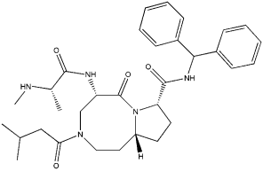
AT-406又称为SM-406、ARRY-334543,是一种有效的、口服生物活性的拟Smac的凋亡蛋白抑制剂(IAP)拮抗剂,与XIAP-BIR3、cIAP1-BIR3和cIAP2-BIR3结合,Ki分别为66.4 nM、1.9 nM和5.1 nM。AT-406可抑制多种癌细胞生长,并诱导小鼠体内移植瘤凋亡。

目前,AT-406已用于临床Phase 1研究阶段。

【该产品仅用于科研实验,不能用于人体】

产品性质

英文别名(English Synonym)

SM-406, ARRY-334543

化学名(Chemical Name)

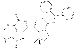
(5S,8S,10aR)-N-(diphenylmethyl)decahydro-5-[[(2S)-2-(methylamino)-1-oxopropyl]amino]-

3-(3-methyl-1-oxobutyl)-6-oxo-pyrrolo[1,2-a][1,5]diazocine-8-carboxamide

靶点(Target)

XIAP

CAS 号(CAS NO.)

1071992-99-8

分子式(Molecular Formula)

C32H43N5O4

分子量(Molecular Weight)

561.71

外观(Appearance)

粉末

纯度(Purity)

≥95%

溶解性(Solubility)

溶于DMSO(100 mg/ml)或乙醇(100 mg/ml)

结构式(Structure)

Smac凋亡蛋白抑制剂|AT-406(SM-406 ARRY-334543) IAP拮抗剂|CAS 1071992-99-8 

运输与保存方法

冰袋运输。粉末直接保存于-20 ºC,有效期2年。易溶于DMSO和乙醇。建议分装后-20ºC避光保存,避免反复冻存,至少可存放6个月。

注意事项

1)为了您的安全和健康,请穿实验服并戴一次性手套操作。

2)粉末溶解前请先短暂离心,以保证产品全在管底。

3)本产品仅用于科研用途,禁止用于人身上。

使用浓度

【具体使用浓度请参考相关文献,并根据自身实验条件(如实验目的,细胞种类,培养特性等)进行摸索和优化。】

相关实验(数据来自于公开发表的文献,仅供参考)

(一)细胞实验(体外研究)

为检测AT-406对癌细胞的作用,用不同剂量AT-406(0-500 μg/ml)处理卵巢癌细胞系,60%卵巢癌细胞对AT-406敏感,IC50<10 μg/ml。另外,AT-406激活细胞凋亡通路,以剂量依赖的方式促进PARP分裂。[2]

参考文献

[1] Cai Q, et al. A Potent and Orally Active Antagonist (SM-406/AT-406) of Multiple Inhibitor of Apoptosis Proteins (IAPs) in Clinical Development for Cancer Treatment. Journal of Medicinal Chemistry 54(8): 2714-2726 (2011).

[2] Brunckhorst MK, et al. AT-406, an orally active antagonist of multiple inhibitor of apoptosis proteins, inhibits progression of human ovarian cancer. Cancer Biol Ther. 13(9):804-811 (2012).

[3] Miura K, et al. Inhibitor of apoptosis protein family as diagnostic markers and therapeutic targets of colorectal cancer. Surg Today. 41(2):175-182 (2011).

HB181128

Smac凋亡蛋白抑制剂|AT-406(SM-406 ARRY-334543) IAP拮抗剂|CAS 1071992-99-8

暂无内容

Smac凋亡蛋白抑制剂|AT-406(SM-406 ARRY-334543) IAP拮抗剂|CAS 1071992-99-8

暂无内容

SM-164 Smac类似物/XIAP拮抗剂/肿瘤细胞凋亡诱导剂|CAS 957135-43-2

SM-164 Smac类似物/XIAP拮抗剂/肿瘤细胞凋亡诱导剂|CAS 957135-43-2

产品说明书

FAQ

COA

已发表文献

产品描述

SM-164是一种有效的细胞通透性Smac类似物,能与XIAP蛋白结合(Ki=0.56 nM),并与cIAP-1和cIAP-2蛋白结合,Ki分别为0.31和1.1 nM。SM-164拮抗XIAP,诱导完全的cIAP-1降解,并诱导TNF-alpha依赖的肿瘤细胞凋亡。在MDA-MB-231移植瘤小鼠中,SM-164有效降解cIAP-1水平,并诱导肿瘤细胞凋亡,引起肿瘤衰退,但对正常小鼠组织没有毒性。

【该产品仅用于科研实验,不能用于人体】

产品性质

英文别名(English Synonym)

SM164

化学名(Chemical Name)

(3S,6R,10aR)-6-((S)-2-(methylamino)propanamido)-N-((R)-(1-(4-(4-(4-(4-((S)-((3S,6S,10aS)-

6-((S)-2-(methylamino)propanamido)-5-oxodecahydropyrrolo[1,2-a]azocine-3-carboxamido)

(phenyl)methyl)-1H-1,2,3-triazol-1-yl)butyl)phenyl)butyl)-1H-1,2,3-triazol-4-yl)

靶点(Target)

IAP

CAS 号(CAS NO.)

957135-43-2

分子式(Molecular Formula)

C62H84N14O6

分子量(Molecular Weight)

1121.42

外观(Appearance)

粉末

纯度(Purity)

>98%

溶解性(Solubility)

溶于DMSO (10 mM)

结构式(Structure)

SM-164 Smac类似物/XIAP拮抗剂/肿瘤细胞凋亡诱导剂|CAS 957135-43-2 

运输与保存方法

冰袋运输。粉末直接保存于-20 ºC,有效期2年。溶于DMSO。建议分装后-20ºC避光保存,避免反复冻存,至少可存放6个月。

注意事项

1)为了您的安全和健康,请穿实验服并戴一次性手套操作。

2)粉末溶解前请先短暂离心,以保证产品全在管底。

3)本产品仅用于科研用途,禁止用于人身上。

使用浓度

【具体使用浓度请参考相关文献,并根据自身实验条件(如实验目的,细胞种类,培养特性等)进行摸索和优化。】

相关实验(数据来自于公开发表的文献,仅供参考)

(一)细胞实验(体外研究)

在SM-164对IAP家族蛋白的亲和力研究中,SM-164作用于XIAP、cIAP-1和cIAP-2蛋白,Ki分别为0.56 nM、0.31和1.1 nM。另外,SM-164剂量依赖性地拮抗XIAP,并诱导caspase活性,诱导细胞凋亡和细胞死亡。[2]

(二)动物实验(体内研究)

在体内实验中,给MDA-MB-231移植瘤小鼠静脉注射5 mg/kg SM-164,SM-164明显降低cIAP-1蛋白水平,而且SM-164诱导明显的细胞凋亡。[2]

参考文献

[1] Zhang S, et al. Smac mimetic SM-164 potentiates APO2L/TRAIL- and doxorubicin-mediated anticancer activity in human hepatocellular carcinoma cells. PLoS One. 7(12): e51461 (2012).

[2] Lu J, et al. SM-164: a novel, bivalent Smac mimetic that induces apoptosis and tumor regression by concurrent removal of the blockade of cIAP-1/2 and XIAP. Cancer Res. 68(22):9384-9393 (2008).

[3] Yang J, et al. Radiosensitization of head and neck squamous cell carcinoma by a SMAC-mimetic compound, SM-164, requires activation of caspases. Mol Cancer Ther. 10(4): 658-669 (2011).

HB181129

SM-164 Smac类似物/XIAP拮抗剂/肿瘤细胞凋亡诱导剂|CAS 957135-43-2

暂无内容

SM-164 Smac类似物/XIAP拮抗剂/肿瘤细胞凋亡诱导剂|CAS 957135-43-2

暂无内容